
THE EASYEF JOURNEY
The workflow at a glance
Choose a stage to explore the screens and details. The system supports decisions; clinicians remain responsible for interpretation.
01
What the workflow needs
The workflow begins with a cardiac ultrasound examination performed by skilled personnel. The phone records the display and sends the clip, so preparation includes equipment, the operator, and the clinician who will review the result.
- An ultrasound machine that can acquire the required cardiac view, and a smartphone that can record the display clearly.
- The EasyEF app and an internet connection for clip submission and result delivery.
- Personnel trained in the workflow, with a clinician to interpret results in the patient’s context.
- An agreed evaluation scope and local data-governance arrangements before a joint evaluation begins.
02
Capture the ultrasound view
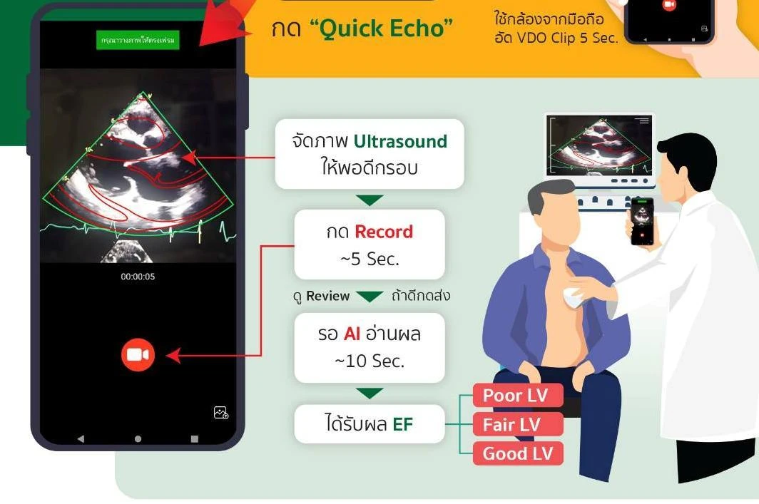
The user records the ultrasound display with a smartphone. The approach evaluated in 2024 used five-second parasternal long-axis clips, showing the heart along its long axis from beside the sternum.
Framing the complete ultrasound region, reducing reflections, and holding the phone steady help produce a suitable input.
References and further reading
03
Check quality before assessment
 Complete image region
 Minimise reflections
 Steady, sufficient recording
Quality screening checks for problems such as reflections, a small or incomplete ultrasound region, multiple image regions, short duration, and excessive movement.
In the 2024 study, 923 of 1,336 submitted clips remained after screening. The study results describe performance on clips meeting those inclusion criteria.
References and further reading
04
Prepare a consistent video input
- 01Identify the ultrasound region
- 02Stabilise the video
- 03Adjust the lighting
Before classification, the pipeline identifies the ultrasound region, stabilizes the video, and adjusts lighting to reduce variation introduced by recording a screen.
The 2024 pipeline used an adapted DAMO-YOLO detector, ORB features for stabilization, and CLAHE with thresholding for lighting adjustment. Each system version needs evaluation tied to its actual pipeline and model.
References and further reading
05
Classify ventricular function
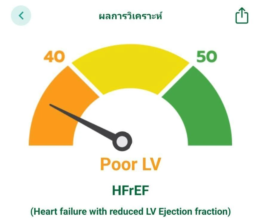
AI analyzes the video sequence to classify left ventricular function. The 2024 system used an adapted ResNet-101, initially predicting five categories before combining them into Reduced EF, Mildly Reduced EF, and Preserved LV.
The output is a functional category rather than a precise continuous EF measurement. Test accuracy was 68.5% for five categories and 96.2% for three, reflecting different levels of classification detail.
References and further reading
06
Return the result to the care team

The clinician brings the information together to make care decisions.
The app submits the clip over the internet to the processing service and displays the returned assessment. The clinician combines the AI output with symptoms, history, and other findings when considering care or further assessment.
The next platform phase connects submission, processing, and a Dashboard / Clinical Report for care teams. Timing evaluation will cover the full journey from recording to result delivery, including network and device conditions.
References and further reading
07
Questions before a service evaluation
- Does it work with every machine? Recording the display reduces reliance on a direct machine connection, but each equipment setup still needs image-quality and compatibility testing.
- Does the phone scan the heart? The ultrasound machine produces the image. The phone records and submits it.
- What does training cover? The planned 1–2-hour session covers the app workflow and clip capture. Cardiac ultrasound acquisition skills require training and supervision appropriate to the user’s role.
References and further reading
08
Extending devices and research questions
Next steps · Development and planned research
We are exploring handheld ultrasound integration and an Echo AI Box concept for existing machines, to study acquisition methods suited to different clinical settings.
Future research questions include valve and pericardial abnormalities. These capabilities need separate development and testing beyond the functional classification already studied.
References and further reading
EasyEF application
For healthcare professionals
Scan a QR code with your phone, or open the store for your device.


SOURCE NOTES
Sources and published project summaries
- 2024 published study — original journal article ↗
- The cardiac-assessment journey — by the EasyEF team
- From an idea to the platform — by the EasyEF team
- EasyEF team: project scope and development roadmap
Project content is compiled and published by the EasyEF team from development records, presentations and study reports. Internal links lead to the team’s summaries, not additional independent evidence. Published research links to its original journal; study status and limitations are stated in the relevant articles.
Explore the source library →